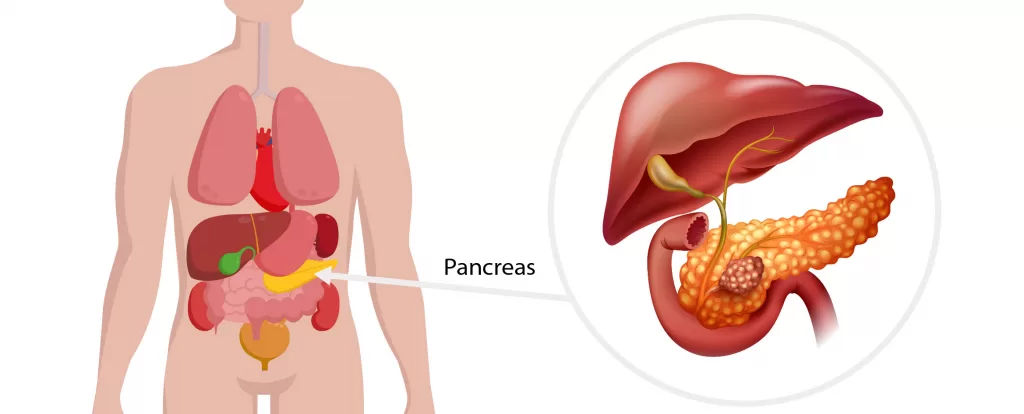
Pancreatic cysts are now commonly diagnosed on CT and ultrasound scans and many are incidentally being found on 80% of abdominal scans undertaken for reasons unrelated to the pancreas. Almost all pancreatic cysts are benign but there is a small but recognised risk of some cysts developing into cancer over a time course of decades. Consequently all pancreatic cysts should be investigated and most are followed for several years. This is usually with MRI scans to monitor growth.
Cysts in the pancreas fall into two categories;
- those that contain saline
- those that contain mucous
Saline containing cysts are benign and may grow during pancreatic development (congenital cysts) or in later life (serous cystadenomas). Saline containing cysts can generally be monitored with imaging but occasionally these are surgically removed if they become large or are causing symptoms.
Mucous containing cysts are associated with a risk of later transformation into cancer.
- Mucinous cystadenomas are benign mucous filled lesions that can grow to moderate size. Generally, these are removed surgically.
- Intraductal papillary mucinous neoplasms (IPMNs) develop when the lining cells of the pancreatic ducts make and secrete mucous. The ducts involved may be very small and at the edge of the pancreas (side branch IPMNs), the main pancreatic duct (main duct IPMNs) or both main and small ducts (mixed IPMNs)
- Main duct IPMNs are rare but do have an elevated risk of cancer and generally pancreatic resection is the treatment of choice. Because the entire pancreatic duct is involved often a total pancreatic resection is necessary to treat these lesions.
Mixed IPMNs also have an elevated risk of developing cancer and are also commonly treated with pancreatic resection. For patients with this condition there is often a period of monitoring growth with regular MRI scans prior to proceeding with surgery. The scans are undertaken specifically to measure the growth (diameter) of the lesions, the development of any solid mass in association with the cyst, and any enlargement of the main pancreatic duct. These three findings are all important in assessing cancer risk. - Side branch IPMNs are common and almost never need treatment. Often the lesions are small (less than 1 cm in diameter) but all are usually monitored for several years with annual MRI scans assessing any growth, change in shape or contour and involvement of the main pancreatic duct.

