INVESTIGATIONS

After you have been seen by a specialist and a history and physical examination has been conducted, sometimes further investigations (tests) may be required in order to gather more information about your condition.

In general, there are three kinds of tests: Scans, Endoscopy and Biopsy. Some tests are a combination of two or all three of these categories.

Please look through the relevant section for information on specific tests that may be ordered in the course of your treatment. You may wish to discuss this further with your specialist and can expect to have the tests and their results explained.

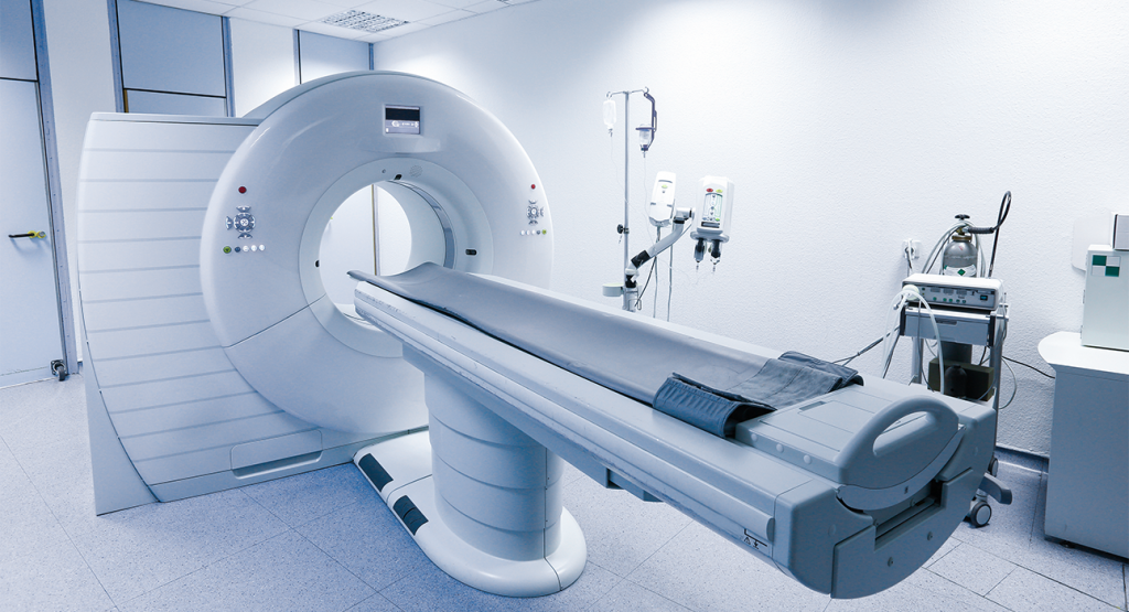
A CT scan (also known as a CAT scan) uses a special machine to take pictures of the inside of your body. The machine takes X-ray images of your body from different angles and uses a computer to combine them into a detailed image.

During the scan, you will lie down on a table that slides into a doughnut-shaped machine. The machine will make a whirring noise as it takes pictures of your body. You will need to stay still while the pictures are being taken, but the scan itself is painless and usually only takes a few minutes. Sometimes the images obtained from a CT scan can be enhanced by the use of contrast, which is a form of medication given via a vein (intravenously) at the time of the scan.

Doctors use CT scans to diagnose and monitor a wide range of medical conditions. They are commonly used for a more detailed view of the area in question- especially for abdominal organs. CT can help establish the presence or absence of a problem or help in diagnosing and staging cancers. They can also help guide medical procedures, such as biopsies or surgeries, by providing detailed images of the affected area. CT scans provide important information and may affect the course of treatment. You should always discuss the specifics of your scan with your specialist.

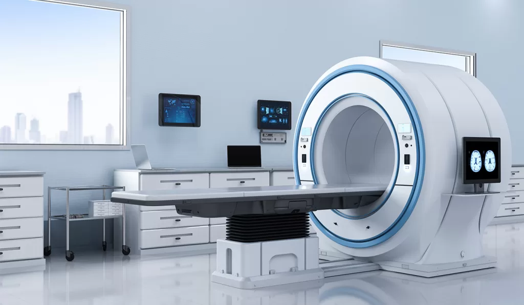
An MRI (Magnetic Resonance Imaging) scan is a medical test that uses a strong magnetic field and radio waves to create detailed pictures of the inside of your body. The machine looks like a big tube and you will need to lie down on a table that slides into the tube.

While you’re inside the tube, the magnetic field causes your body’s atoms to move in a certain way. Then, radio waves are sent through your body, and these atoms emit signals that are picked up by a special computer. The computer then uses this information to create a detailed picture of the inside of your body.

The process is completely painless, but it can be a little noisy as the machine makes some loud banging and knocking sounds during the scan. You will need to lie very still during the scan, which can take around 30 to 60 minutes.

The images produced by the MRI scan can help doctors to diagnose and treat a variety of medical conditions, such as tumours, injuries, and diseases affecting the body. MRI is often used in conjunction with CT scans and offers greater detail for very specific questions. In most circumstances, a CT scan is adequate to answer the specific clinical question but sometimes an MRI is used in addition. E.g. to determine the presence of liver lesions not clearly seen on CT.

A CT-PET scan is a type of medical imaging test that combines two different technologies: computed tomography (CT) and positron emission tomography (PET).

During a CT scan, a series of X-ray images are taken from different angles around your body to create a detailed 3D image. This allows doctors to see your organs and tissues in great detail.

A PET scan uses a special radioactive substance called a tracer, which is injected into the body. The tracer travels to the organs and tissues being examined and emits tiny particles called positrons. The PET scanner detects these positrons and creates an image of the activity in your body. In many cases, it is radiolabelled glucose that is given and those tissues which are taking up high levels of glucose light up more brightly on the scan. Therefore, your specialist will have both the images normally obtained from a CT plus a superimposed metabolic picture. Since tissues that are using glucose rapidly include cancers or infection, these areas show up brightly allowing for interpretation.

When CT and PET scans are combined, doctors can get both anatomical and functional information about your body. This can be helpful in diagnosing some types of cancer as well as other diseases. There are very specific indications in which CT-PET scans are used (oesophageal cancer; advanced melanoma) and others where they are used infrequently (e.g. pancreas cancer) Your specialist can discuss the role of CT-PET scans in your treatment.

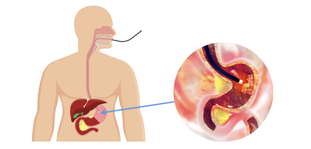
Endoscopy is a medical procedure that allows a doctor to examine the inside of the body using a flexible tube with a tiny camera attached to the end of it. The tube, called an endoscope, is inserted into the body through a natural opening, such as the mouth, nose, or anus, or through a small incision. Endoscopy is a semi-invasive procedure. An Upper GI Endoscopy (Gastroscopy) is when a fibre-optic telescope is inserted under sedation from the mouth to look at the mouth, the oesophagus, the stomach and part of the small intestine. A colonoscopy is when a fibreoptic telescope is introduced under sedation from the anus to look at the anus, rectum, entire colon and the last part of the small intestine.

As the endoscope is moved through the body, the camera sends images to a monitor, allowing the doctor to see and assess the condition of organs, tissues, and structures inside the body. Depending on the type of endoscopy, the doctor may also be able to take samples of tissue for further examination, or perform certain medical treatments through the endoscope. Sometimes, just looking via endoscopy is enough to make a diagnosis (for example-a hiatus hernia). Biopsies can also be taken during endoscopy to help diagnose a problem and in select circumstances, treatment can be accomplished such as resection of an early stomach cancer.

Endoscopy is commonly used to diagnose and treat conditions of the digestive tract, such as ulcers, inflammation, and cancers. Your specialist can discuss the role of endoscopy in your care both from a diagnostic and therapeutic perspective.

A diagnostic laparoscopy is a surgical procedure used to visualize the organs inside the abdomen and pelvis. It is a minimally invasive technique that involves making a small incision in the abdomen and inserting a thin, flexible tube with a camera attached to it, called a laparoscope. This is usually done under a general anaesthetic and permits the surgeon to visualize the organs directly and/or take biopsies or proceed with curative surgery if appropriate. There is an established but limited role for diagnostic laparoscopy as usually, less invasive means of investigation are considered first or alongside this technique.

During a diagnostic laparoscopy, the surgeon can examine the organs in the abdomen and pelvis, such as the uterus, ovaries, fallopian tubes, bladder, and intestines. The laparoscope provides a clear view of these organs, allowing the surgeon to identify any abnormalities or conditions that may be causing symptoms such as pain.

If an abnormality is found, the surgeon may take a tissue sample (biopsy) for further testing or perform additional procedures to treat the problem. Most people can go home the same day or the day after the procedure.

Ultrasound is a diagnostic imaging technique that uses high-frequency sound waves to create images of organs and tissues inside the body.

During an ultrasound exam, a small handheld device called a transducer is placed on the skin and it emits sound waves into the body. These sound waves bounce off the organs and tissues inside the body and are detected by the same transducer. The returning sound waves are used to create images of the internal structures, which can be seen on a screen and interpreted by a trained medical professional. Ultrasound imaging is non-invasive, safe, and does not use ionizing radiation, making it a valuable tool in diagnosing a wide range of medical conditions. It is also used in pregnancy for this reason.

Ultrasound is commonly used to diagnose gallstones but can also be used to look at the other organs in the abdomen.


An endoscopic ultrasound (EUS) is a minimally invasive diagnostic test that combines endoscopy and ultrasound imaging. During the procedure, an endoscope (a flexible tube with a light and camera on the end) is passed through the mouth or anus and into the digestive tract. At the end of the endoscope is an ultrasound probe that emits high-frequency sound waves to create detailed images of the internal organs and tissues surrounding the digestive tract. The premise of endoscopic ultrasound is to get superior quality ultrasound pictures by having the probe closer to the actual organs themselves whilst being minimally invasive.

Endoscopic ultrasound is commonly used to evaluate and diagnose conditions affecting the digestive tract, such as:

  1. Abdominal pain
  2. Chronic pancreatitis
  3. Gallstones
  4. Gastrointestinal tumours
  5. Lymphoma
  6. Rectal cancer
  7. Staging of oesophageal cancer
  8. Staging of pancreatic cancer

The scope uses its position in the stomach or intestine (as appropriate) to obtain excellent views of the regional organs such as the liver, gallbladder and pancreas. It also allows for safe biopsy of the pancreas and may provide important technical information if surgery of the pancreas is being considered.

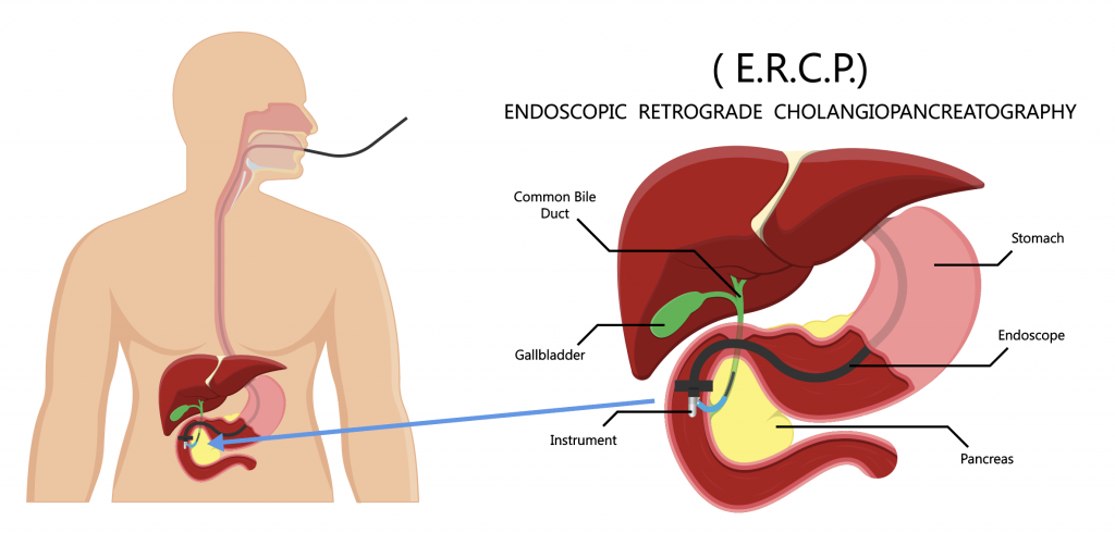
ERCP stands for Endoscopic Retrograde Cholangiopancreatography. It is a medical procedure that combines endoscopy and X-ray imaging to examine and treat conditions affecting the bile ducts, pancreas, and gallbladder.

During an ERCP, an endoscope is inserted through the mouth, down the throat, and into the stomach and duodenum (the first part of the small intestine). A small tube is then passed through the endoscope and into the bile and pancreatic ducts. Contrast dye is injected through the tube, and X-ray images are taken to visualize the structures and identify any abnormalities.

ERCP can be used to diagnose and treat conditions such as gallstones, blockages in the bile ducts or pancreatic ducts, inflammation of the pancreas, and cancers of the bile ducts or pancreas. It can also be used to remove gallstones, take tissue samples for biopsy, or place stents to open up narrowed or blocked ducts.

In many instances, the diagnostic role of ERCP has been replaced by specialized MRI scans but ERCP remains an important therapeutic test. Please note that ERCP is accompanied by specific risks such as those of bleeding, intestinal perforation and pancreatitis and your specialist endoscopist will be able to discuss these with you before the procedure.

Percutaneous transhepatic cholangiography (PTC) is a medical procedure used to examine the bile ducts within the liver. During the procedure, a needle is inserted through the skin and into the liver to access the bile ducts. A contrast dye is then injected through the needle, and X-ray images are taken to visualize the bile ducts.

PTC is often used to diagnose and treat problems such as blockages or narrowing in the bile ducts, tumours, and gallstones. It can also be used to place a stent, a small tube, into the bile duct to help keep it open and allow bile to flow freely. The procedure is usually performed under local anaesthesia and may require a short hospital stay.

This test is often used in the context of complex cancer of the liver, pancreas and bile ducts. It is performed by a specialist radiologist and is accompanied by risks such as bleeding, perforation and bile leak. If this test is recommended by your specialist, you can expect to have the indications and the risks specifically discussed with you.

Liver biopsy means to take a small sample of the liver for analysis under the microscope (histological analysis). It is sometimes indicated in the context of intrinsic diseases of the liver (e.g. cirrhosis) or for suspicious lesions where cancer is suspected. Liver biopsy can be done either percutaneously (through the skin under imaging guidance) or during surgery.

Portal vein embolization (PVE) and Hepatic Vein Embolization (HVE) are medical procedures used to treat patients who require a major liver resection. The procedure involves blocking or partially blocking the portal vein, which is the main blood vessel that brings blood to the liver.

During PVE, a radiologist will insert a catheter through a vein in your groin and guide it to the portal vein in your liver. Once the catheter is in place, the radiologist will inject a substance (such as coils, particles, or other agents) into the vein to block the flow of blood to a specific part of the liver.

The purpose of PVE is to redirect blood flow away from the portion of the liver that will be removed during the liver resection. It can help deprive blood flow to one side of the liver (for example a side with cancer in it) and promote growth in the other side of the liver. It can be an important part of a cancer control strategy for primary and secondary cancers of the liver. This allows the remaining portion of the liver to grow and compensate for the loss of function of the removed part. PVE is usually performed several weeks before the liver resection to allow the liver to regenerate.

PVE is generally considered a safe and effective procedure, but like any medical procedure, it does carry some risks. These may include bleeding, infection, and damage to other organs. Your doctor will discuss the risks and benefits of PVE with you before recommending the procedure. If your specialist recommends this treatment to you as a potential precursor to surgery, you can expect to have the risks and indications discussed with you.